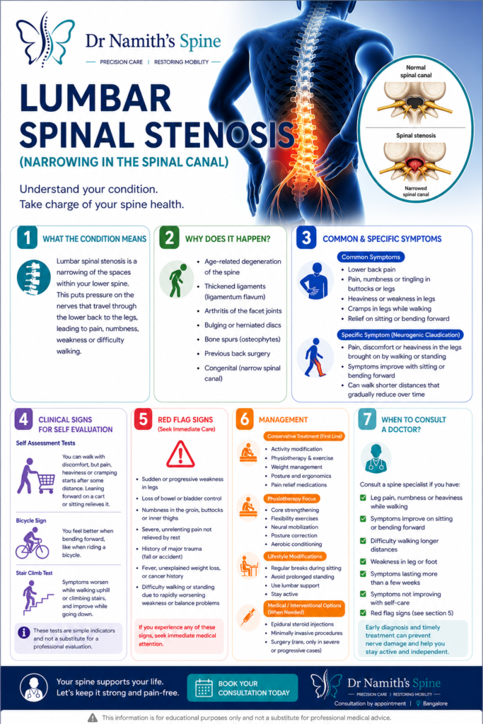
What is Lumbar Spinal Stenosis?
Lumbar spinal stenosis is a narrowing of the spinal canal in the lower back, resulting in compression of the nerve roots within. Unlike a disc prolapse where compression tends to be acute and at a single level, lumbar stenosis is typically a gradual, degenerative process affecting one or more levels and is particularly common in adults over 60.
The condition is one of the most common reasons for spine surgery in older adults worldwide, and while it can significantly limit quality of life, it is very effectively treated when managed appropriately.
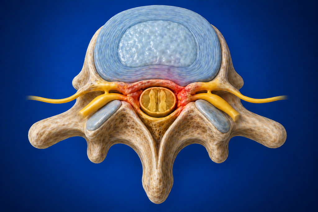
Who Gets It and Why?
Lumbar spinal stenosis develops as a result of age-related changes that collectively narrow the spinal canal:
- Disc degeneration and bulging
- Facet joint arthritis and enlargement (hypertrophy)
- Thickening of the ligamentum flavum
- Bony spurs (osteophytes) encroaching on the canal
- Spondylolisthesis (forward slip of one vertebra on another) contributing to dynamic narrowing
Congenital stenosis, where the canal is naturally narrow from birth, means that degenerative changes produce symptoms earlier and more severely than in a normally sized canal.
What Does It Feel Like?
The classic presentation of lumbar spinal stenosis is neurogenic claudication, which is a highly characteristic symptom pattern:
- Pain, cramping, heaviness, or numbness in the legs that comes on with walking or standing
- Symptoms typically affect both legs, though may be worse on one side
- Relief on sitting down, bending forward (such as leaning on a shopping trolley), or lying down with knees bent
- Patients often describe being able to walk only a limited distance before needing to stop and rest
- Cycling is typically well tolerated because the flexed posture opens the spinal canal
This pattern distinguishes neurogenic claudication from vascular claudication (poor blood supply to the legs), where rest relieves symptoms regardless of position.
Back pain is commonly associated, though leg symptoms usually dominate the clinical picture.
How Is It Diagnosed?
- MRI lumbar spine is the primary investigation, demonstrating the level and severity of canal narrowing and nerve root compression
- CT scan for detailed bony anatomy and when surgical planning requires precise assessment of the facet joints and bony canal
- Vascular assessment to exclude peripheral arterial disease when claudication symptoms are present
- Standing X-rays to assess spinal alignment and detect spondylolisthesis
How Is It Treated?
Mild to moderate stenosis without significant neurological deficit is initially managed non-surgically:
- Physiotherapy with lumbar flexion-based exercises, core strengthening, and posture training
- Pain management including anti-inflammatory medications and nerve pain agents
- Epidural steroid injections providing meaningful relief for selected patients
- Activity modification and walking aids if helpful
Surgery is recommended when quality of life is significantly impaired despite adequate non-surgical treatment, when neurological deficits are present or worsening, or when bladder and bowel function is threatened.
Lumbar decompression (laminectomy or laminotomy) removes the tissue compressing the nerve roots and is one of the most successful operations in spine surgery. Where instability coexists, decompression with spinal fusion is performed. Minimally invasive techniques are used wherever feasible to reduce blood loss, post-operative pain, and recovery time.
The large majority of patients experience significant improvement in walking ability and leg pain following surgery.
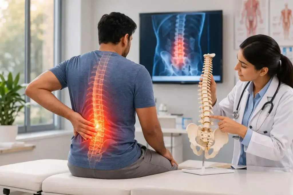
Self-Check at Home
The Shopping Trolley Test: This is one of the most practical self-checks for lumbar spinal stenosis. The next time you are in a supermarket, notice whether leaning forward on the shopping trolley significantly relieves your leg pain and allows you to walk further than you can without it. This forward-flexed posture opens the narrowed spinal canal and reduces nerve compression — a posture-dependent symptom pattern that is highly characteristic of lumbar spinal stenosis.
The Bicycle Test: Can you ride a stationary or regular bicycle comfortably for longer than you can walk? The flexed-forward seated posture on a bicycle opens the lumbar canal, allowing nerve roots more space. Patients with lumbar stenosis typically tolerate cycling far better than walking — the opposite of what is seen in vascular claudication (poor blood supply), where cycling causes equal symptoms to walking.
The Walking Distance and Recovery Time Test: Walk at your normal pace and note:
- How far can you walk before leg symptoms (pain, cramp, heaviness, numbness) force you to stop?
- How long do you need to rest before you can walk again?
- Does sitting or bending forward speed up your recovery compared to standing still?
Progressive shortening of your walking distance over months, combined with rapid relief on sitting, is the hallmark of neurogenic claudication from lumbar stenosis.
Unsure about your symptoms? Our free Spine Self Check takes less than two minutes and gives you a clearer picture before your consultation.
Motion-Preserving Surgical Options
Minimally Invasive Decompression (Laminotomy / Unilateral Approach Bilateral Decompression – ULBD): When surgery is needed for lumbar stenosis, the motion-preserving approach is decompression without fusion wherever possible. Modern minimally invasive techniques, including the unilateral approach bilateral decompression (ULBD), allow the surgeon to decompress both sides of the spinal canal through a single small incision on one side, using a tubular or endoscopic retractor. This preserves the facet joints, interspinous ligaments, and contralateral muscles — the structures that maintain spinal stability and movement.
Why Avoid Unnecessary Fusion in Stenosis: Fusion for lumbar stenosis is appropriate when instability or spondylolisthesis coexists, but adding fusion purely for decompression of stable stenosis increases surgical risk, recovery time, blood loss, and permanently reduces lumbar mobility. The evidence supports decompression alone as equally effective to fusion for stable stenosis, and this is the approach followed at this practice.
Understanding your symptoms is the first step. If something here sounds familiar, book a consultation with Dr. Namith Rangaswamy or call +91 91081 04114 to get a proper diagnosis.