
What is Sciatica?
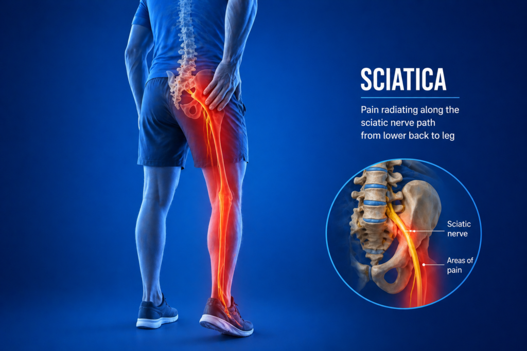
Sciatica is the term used to describe pain that travels along the path of the sciatic nerve, which runs from the lower back through the buttock and down the back of each leg. It is not a diagnosis in itself, but a symptom of an underlying condition compressing or irritating one of the nerve roots that form the sciatic nerve.
It is one of the most common reasons people visit a spine specialist, and while it can be severely debilitating, the majority of patients recover well with the right treatment approach.
Who Gets It and Why?
The most common underlying causes include:
- Lumbar disc herniation (prolapsed disc) pressing on a nerve root, the most frequent cause in younger and middle-aged adults
- Lumbar spinal stenosis causing nerve root compression, more common in older adults
- Foraminal stenosis where the bony canal through which the nerve exits the spine becomes narrowed
- Piriformis syndrome where the sciatic nerve is compressed by a muscle in the buttock, a less common but important cause
- Spondylolisthesis where one vertebra slips forward on another, compressing the nerve
What Does It Feel Like?
The hallmark of sciatica is pain that starts in the lower back or buttock and travels down the leg, often reaching the calf or foot. The character of the pain is typically:
- Sharp, burning, or electric shock-like
- Shooting down one leg, following a specific pathway depending on which nerve is affected
- Worsened by sitting, coughing, sneezing, or straining
- Sometimes accompanied by numbness or tingling in the leg or foot
- In more significant cases, weakness in the leg or foot (foot drop in severe cases)
Pain affecting both legs simultaneously, or associated with bladder or bowel dysfunction, is a red flag that requires urgent evaluation.
How Is It Diagnosed?
- Clinical examination including straight leg raise test and neurological assessment to identify the affected nerve level
- MRI of the lumbar spine is the investigation of choice, identifying the cause and level of nerve compression
- CT scan when MRI is unavailable or for additional bony detail
- Nerve conduction studies in selected cases to confirm nerve involvement
How Is It Treated?
Most cases of sciatica improve with time and non-surgical management:
- Physiotherapy focusing on core strengthening, nerve mobilisation, and posture correction
- Anti-inflammatory medications and nerve pain agents for symptom control
- Transforaminal epidural steroid injections providing targeted relief, particularly useful for acute flare-ups
- Activity modification during the acute phase, avoiding prolonged sitting
Surgery is indicated when neurological deficits are progressive, when pain is severe and persistent despite adequate non-surgical treatment (typically 6 to 12 weeks), or in the case of cauda equina syndrome, which is a surgical emergency. Minimally invasive microdiscectomy or endoscopic discectomy achieves excellent results for appropriate candidates, with most patients returning to normal activity within weeks.
Sciatica is very treatable. Early specialist assessment ensures the right diagnosis and avoids unnecessary delay in recovery.
Self-Check at Home
The Straight Leg Raise Test (SLR): Lie flat on your back on a firm surface. With your knee straight, have someone slowly raise your leg by lifting the heel upward. Alternatively, do this yourself by lifting your leg while keeping the knee fully extended. Note the angle at which pain begins.
Pain shooting down the back of the leg (not just local back or hip pain) when the leg is raised between 30 and 70 degrees is a positive SLR — one of the most reliable clinical tests for lumbar nerve root compression from a disc herniation. Pain only at very high angles (above 70 degrees) is less specific.
The Crossed SLR: While lying flat, raise the leg on the side without symptoms. If this reproduces sciatic pain in the opposite (symptomatic) leg, it is a highly specific sign of significant nerve root compression, often from a large central or paracentral disc herniation.
The Dermatomal Sensory Check: Compare sensation (light touch) on both legs:
- Outer calf and top of foot: L5 nerve root
- Heel and outer border of foot: S1 nerve root
Reduced sensation in a specific area compared to the other side helps localise the affected nerve level.
The Muscle Strength Check:
- Walk on your heels (lift your toes off the floor while walking). Difficulty or asymmetry suggests L4-L5 nerve involvement (tibialis anterior weakness).
- Walk on your tiptoes. Difficulty or asymmetry suggests L5-S1 nerve involvement (gastrocnemius weakness).
Unsure about your symptoms? Our free Spine Self Check takes less than two minutes and gives you a clearer picture before your consultation.
Motion-Preserving Surgical Options
Microdiscectomy and Endoscopic Discectomy: When surgery is needed for sciatica from disc herniation, the goal is always to remove only the herniated fragment causing compression while leaving the disc itself intact wherever possible. This preserves disc height, segmental mobility, and natural shock-absorbing function.
Endoscopic Discectomy: Full-endoscopic lumbar discectomy is the most minimally invasive surgical option available for lumbar disc herniation causing sciatica. Performed through a small tube using a camera and specialised instruments, it removes the herniated disc fragment through a skin incision of less than 1 cm, with minimal muscle disruption, minimal blood loss, and rapid return to activity. Many patients are discharged the same day or the following morning.
This approach exemplifies the motion-preserving, tissue-sparing philosophy applied throughout this practice.
Learn more: What is sciatica really, and why does it cause leg pain when the problem is in your back?
Understanding your symptoms is the first step. If something here sounds familiar, book a consultation with Dr. Namith Rangaswamy or call +91 91081 04114 to get a proper diagnosis.