
What is a Prolapsed Intervertebral Disc?
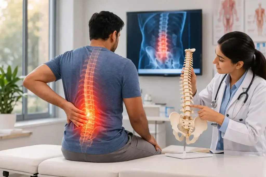
Each vertebra in the spine is separated by an intervertebral disc, which acts as a shock absorber and allows movement between spinal segments. Each disc has a tough outer ring (annulus fibrosus) and a soft, gel-like centre (nucleus pulposus). A prolapsed intervertebral disc, also called a herniated or slipped disc, occurs when the soft inner material pushes through a tear in the outer ring and presses on nearby nerve roots or, in severe cases, the spinal cord or cauda equina.
Lumbar disc prolapse most commonly occurs at the L4-L5 or L5-S1 levels and is one of the most frequent causes of back and leg pain in adults between the ages of 30 and 55.
Who Gets It and Why?
- Age-related disc degeneration weakens the annulus, making it more vulnerable to tearing
- Heavy lifting with poor technique, particularly with a flexed and rotated spine
- Prolonged sitting and sedentary lifestyle increasing disc load
- Obesity increasing mechanical stress on lumbar discs
- Genetic predisposition to early disc degeneration
- Occasionally a specific incident such as a fall or sudden strain, though many occur without a clear trigger
What Does It Feel Like?
Symptoms depend on the size and direction of the prolapse and which nerve is affected:
- Low back pain, which may precede leg symptoms
- Leg pain (sciatica) following the distribution of the compressed nerve root
- Numbness or tingling in the leg, calf, or foot
- Weakness in specific leg muscles depending on the nerve level involved
- Worsening of symptoms with sitting, bending forward, coughing, or straining
- Relief on lying down in a comfortable position
A large central disc prolapse affecting multiple nerve roots simultaneously can cause cauda equina syndrome, which is a surgical emergency (see separate page).
How Is It Diagnosed?
- MRI lumbar spine is the gold standard investigation, clearly showing the disc prolapse, its direction, and the degree of nerve compression
- CT scan when MRI is contraindicated or for pre-surgical planning
- Clinical correlation is essential, as MRI findings must match the patient’s symptoms and neurological examination for accurate treatment planning
How Is It Treated?
The natural history of lumbar disc prolapse is favourable in the majority of cases, with significant improvement over 6 to 12 weeks in many patients:
- Physiotherapy with McKenzie method exercises, core strengthening, and nerve mobilisation
- Oral medications including anti-inflammatories, muscle relaxants, and nerve pain agents
- Transforaminal epidural steroid injections for targeted relief and to facilitate rehabilitation
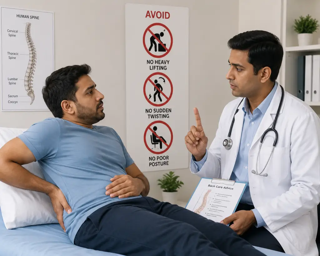
- Lifestyle modification including weight management, ergonomic improvements, and activity guidance
Surgical intervention is considered for:
- Progressive or severe neurological deficit (weakness, significant numbness)
- Cauda equina syndrome (emergency)
- Persistent disabling pain after 6 to 12 weeks of structured non-surgical treatment
Microdiscectomy (microscope-assisted minimally invasive removal of the prolapsed disc fragment) and Endoscopic discectomy (keyhole surgery through a small tube) are the standard surgical procedures. Both offer excellent outcomes with low complication rates and rapid recovery. The choice between them is guided by the patient’s anatomy, prolapse type, and surgeon expertise.
A prolapsed disc does not automatically mean surgery. A structured, evidence-based approach gives the majority of patients a full recovery without the need for an operation.
Self-Check at Home
The SLR Test: Perform the straight leg raise as described in the sciatica section above. A positive SLR in the context of back and leg pain is strongly suggestive of lumbar disc prolapse with nerve root compression.
The Valsalva Check: Note whether your leg pain or back pain worsens when you cough, sneeze, or strain for a bowel movement. Worsening of radicular (leg) pain with these activities reflects increased intradiscal and intraspinal pressure momentarily compressing an already irritated nerve root — a characteristic feature of disc prolapse.
The Sitting vs Standing Check:
- Does sitting for more than 20 to 30 minutes significantly worsen your leg pain?
- Does standing and walking also aggravate symptoms but to a lesser degree than sitting?
- Is lying down in a specific position (often on the side with knees bent) the most comfortable?
This pattern — sitting worst, lying down best — is typical of a disc herniation with nerve root irritation, as sitting maximises intradiscal pressure.
Unsure about your symptoms? Our free Spine Self Check takes less than two minutes and gives you a clearer picture before your consultation.
Motion-Preserving Surgical Options
Endoscopic Discectomy: The preferred surgical approach for lumbar disc prolapse at this practice. A full-endoscopic technique removes only the prolapsed fragment under direct visualisation through a pencil-sized tube, leaving the rest of the disc, the facet joints, and all surrounding muscles completely undisturbed. This is the ultimate expression of motion preservation in lumbar disc surgery — treating the problem without altering the mechanics of the spine at all.
Why Not Fusion for a Disc Prolapse? Disc prolapse treated surgically does not require fusion in the vast majority of cases. Fusion adds unnecessary motion loss, longer surgery, higher complication rates, and greater recovery burden when simple fragment removal achieves the same outcome. Fusion for lumbar disc prolapse is reserved for rare cases where associated instability or deformity coexists.
Understanding your symptoms is the first step. If something here sounds familiar, book a consultation with Dr. Namith Rangaswamy or call +91 91081 04114 to get a proper diagnosis.