
What is Foraminal Stenosis?
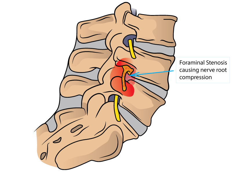
Foraminal stenosis refers to narrowing of the foramen, the bony tunnel through which each spinal nerve root exits the spinal canal on its way to the arms or legs. When this tunnel becomes constricted, the nerve root passing through it is compressed, producing pain, numbness, or weakness in the limb it supplies.
While spinal stenosis refers to narrowing of the central canal, foraminal stenosis specifically affects the lateral exit points for individual nerve roots. It can occur in the cervical, thoracic, or lumbar spine, but is most clinically significant in the lumbar and cervical regions.
Who Gets It and Why?
Foraminal stenosis develops as a result of:
- Disc degeneration and collapse reducing foraminal height as the disc space narrows
- Facet joint arthritis with bony overgrowth encroaching on the foramen from behind
- Disc herniation into the foramen (far lateral disc herniation)
- Spondylolisthesis causing foraminal distortion as one vertebra shifts on another
- Post-surgical scarring in patients who have had previous spine surgery
It is an important cause of persistent or recurrent leg pain after previous lumbar surgery, which may indicate adjacent segment disease or post-surgical foraminal narrowing rather than recurrent disc prolapse.
What Does It Feel Like?
- Unilateral leg pain (one side) following the distribution of the compressed nerve root
- Numbness or tingling in a specific part of the leg or foot
- Weakness in specific muscle groups supplied by the affected nerve
- Pain often worsened by standing upright or extending the spine (leaning back), which further narrows the foramen
- Relief on flexing forward or sitting
Foraminal stenosis at L5 classically causes pain and numbness along the outer leg and top of the foot. At S1, symptoms travel down the back of the leg into the heel and outer border of the foot.
How Is It Diagnosed?
- MRI lumbar spine with attention to the foraminal zones at each level
- CT scan for detailed assessment of bony foraminal anatomy
- Selective nerve root injection serves a dual diagnostic and therapeutic purpose, confirming that the targeted nerve root is the source of symptoms while providing relief
- Clinical neurological examination to correlate the level of compression with the patient’s symptom distribution
How Is It Treated?
Non-surgical treatment forms the first line of management:
- Physiotherapy with lumbar extension-avoidance exercises and nerve mobilisation
- Analgesic and anti-inflammatory medications
- Transforaminal selective nerve root injections are particularly effective for foraminal stenosis, delivering steroid precisely to the affected nerve root
Surgical decompression is indicated for persistent or progressive neurological deficit. The standard procedure is foraminotomy, enlarging the bony foramen to free the compressed nerve root. This can be performed using minimally invasive or endoscopic techniques, allowing a targeted decompression with minimal disruption to surrounding structures. Where instability is present, fusion may be combined with decompression.
Self-Check at Home
The Extension Provocation Check: Stand upright and gently lean backward (extend your lumbar spine). If this reproduces or significantly worsens your leg pain or numbness, it suggests foraminal stenosis — extension narrows the foramen further, increasing nerve root compression.
The Flexion Relief Check: Sit down or bend slightly forward from the standing position. If this promptly relieves your leg symptoms, it confirms the positional nature of foraminal compression, as flexion opens the foramen and reduces nerve pressure.
The Single Level Symptom Pattern Check: Foraminal stenosis typically causes very specific, consistent symptoms following a single nerve root distribution:
- Pain and tingling in the outer shin and top of the foot: L5 foraminal stenosis
- Pain in the back of the thigh, calf, and outer border of the foot: S1 foraminal stenosis
A highly consistent, reproducible symptom pattern in a specific nerve distribution, worsened by standing and relieved by sitting, is characteristic of foraminal nerve root entrapment.
Motion-Preserving Surgical Options
Endoscopic Foraminotomy: Full-endoscopic transforaminal or interlaminar foraminotomy is the most minimally invasive approach to foraminal decompression available. Using a small-diameter endoscope placed directly at the foramen, the compressing bone and ligament are removed under direct vision through a skin incision of under 1 cm. The disc, facet joint, and stabilising ligaments are preserved, maintaining complete segmental mobility.
This is the preferred surgical approach for isolated foraminal stenosis at this practice, offering same-day or next-day discharge, minimal postoperative pain, and rapid return to activity.
Understanding your symptoms is the first step. If something here sounds familiar, book a consultation with Dr. Namith Rangaswamy or call +91 91081 04114 to get a proper diagnosis.