
What is Cervical Disc Herniation?
A cervical disc herniation occurs when the soft inner core of an intervertebral disc in the neck ruptures through its tough outer layer and presses on an adjacent nerve root or, in more serious cases, the spinal cord itself. It is one of the most common causes of neck and arm pain encountered in a spine clinic, and while it can be severely debilitating, the majority of patients recover well with appropriate management.
Each disc in the cervical spine sits between two vertebrae, acting as a shock absorber and allowing controlled movement. The disc has two components — a tough outer ring called the annulus fibrosus and a soft, gel-like centre called the nucleus pulposus. When the annulus develops a tear, the nucleus can herniate through it, compressing the nearby nerve root (causing radiculopathy) or the spinal cord (causing myelopathy).
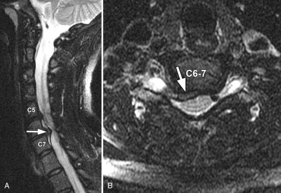
Cervical disc herniation most commonly occurs at C5-C6 and C6-C7, the most mobile and mechanically stressed levels of the cervical spine.
Who Gets It and Why?
- Age-related disc degeneration weakening the annulus fibrosus, making it vulnerable to tearing
- Acute trauma including whiplash, falls, or direct impact
- Repetitive mechanical stress from prolonged desk work, driving, or heavy manual labour
- Sudden forceful neck movements
- Genetic predisposition to early disc degeneration
- Smoking, which accelerates disc degeneration by impairing disc nutrition
Unlike lumbar disc herniation, which is strongly associated with bending and lifting, cervical disc herniation often occurs without a clear precipitating event.
What Does It Feel Like?
Symptoms depend on whether the herniation compresses a nerve root, the spinal cord, or both:
Nerve root compression (radiculopathy):
- Sharp, burning, or electric shock-like pain radiating from the neck into the shoulder, arm, forearm, or hand
- Numbness or tingling in specific fingers depending on the nerve level affected
- Weakness in specific arm or hand muscles
- Symptoms worsened by turning the head, looking up, or coughing
Spinal cord compression (myelopathy):
- Hand clumsiness and difficulty with fine motor tasks
- Weakness or heaviness in both arms and legs
- Unsteady walking
- Bladder dysfunction in advanced cases
Some patients experience both radiculopathy and myelopathy simultaneously (radiculomyelopathy), with arm pain and hand weakness alongside walking difficulty.

Self-Check at Home
The Spurling’s Test (Home Version): Sit upright on a firm chair. Tilt your head toward the side of your arm pain and gently apply light downward pressure on top of your head with your hand. If this reproduces or worsens the shooting pain, numbness, or tingling down your arm, it suggests nerve root compression from a cervical disc herniation. Perform this gently and stop immediately if significant pain occurs.
The Shoulder Abduction Relief Test: Slowly raise the arm on your symptomatic side and rest your hand on top of your head. If this position relieves your arm pain or tingling, it is a positive shoulder abduction sign — a characteristic feature of cervical nerve root compression. The movement reduces tension on the compressed nerve root.
The Finger Numbness Map: Using light touch, compare sensation in both hands finger by finger:
- Thumb and index finger numbness: C6 nerve root, most commonly from C5-C6 disc herniation
- Middle finger numbness: C7 nerve root, most commonly from C6-C7 disc herniation
- Ring and little finger numbness: C8 nerve root, most commonly from C7-T1 disc herniation
Consistent numbness in a specific finger or fingers on one side, combined with neck and arm pain, is a reliable indicator of the affected disc level.
The Hand Function Check: Open and close your fist as quickly as possible for 10 seconds and count the cycles. Fewer than 20 cycles, particularly combined with arm pain and neck symptoms, suggests possible spinal cord involvement from a large central disc herniation. Seek specialist evaluation promptly.
Unsure about your symptoms? Our free Spine Self Check takes less than two minutes and gives you a clearer picture before your consultation.
How Is It Diagnosed?
- MRI of the cervical spine is the investigation of choice, clearly showing the disc herniation, its direction, and the degree of nerve root or cord compression
- CT scan for additional bony detail, particularly in older patients where concurrent spondylotic changes need precise characterisation
- Nerve conduction studies and EMG to confirm nerve involvement and localise the level when clinical and imaging findings need further correlation
- Dynamic X-rays to assess cervical alignment and any instability at the affected level
How Is It Treated?
The natural history of cervical disc herniation is favourable in many patients, particularly those with soft herniations causing radiculopathy:
- Physiotherapy with cervical traction, nerve mobilisation, and postural correction
- Anti-inflammatory and nerve pain medications
- Selective nerve root injections for targeted relief and to facilitate rehabilitation
- Activity modification during the acute phase
Surgery is indicated for progressive neurological deficit, myelopathy from cord compression, or persistent disabling symptoms after adequate non-surgical treatment.
Motion-Preserving Surgical Options
Cervical Disc Replacement (Artificial Disc Surgery)
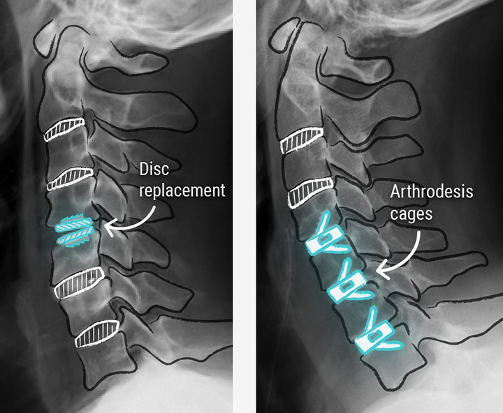
Cervical disc replacement is the premier motion-preserving surgical treatment for cervical disc herniation and is the preferred surgical approach at this practice for appropriate candidates.
The herniated disc is removed through a small anterior neck incision, the nerve root or spinal cord is decompressed, and an artificial disc implant is placed in the disc space. The implant replicates the natural disc’s function — maintaining the disc space height, allowing continued movement at the treated level, and preserving the natural cervical range of motion.
Advantages over fusion:
- Normal cervical movement is maintained at the treated level
- The risk of adjacent segment disease (accelerated degeneration at levels above and below) is significantly reduced
- Return to normal activities including sport and physically demanding work is faster
- No need for bone grafting or cage insertion
- Multiple high-quality randomised controlled trials demonstrate equivalent or superior outcomes to fusion at 7 to 10 year follow-up
Who is suitable: Disc replacement is ideally suited to patients with one or two level cervical disc herniation causing radiculopathy or early myelopathy, preserved cervical alignment, no significant facet joint arthritis, and good bone density. The treating surgeon will assess suitability based on imaging and clinical findings.
Why This Is the Practice Differentiator: Cervical disc replacement requires specific surgical training and implant expertise. At this practice, disc replacement is offered as the standard surgical option for eligible patients rather than as an exception, reflecting a fundamental commitment to preserving the patient’s natural spinal movement wherever surgically possible.
Understanding your symptoms is the first step. If something here sounds familiar, book a consultation with Dr. Namith Rangaswamy or call +91 91081 04114 to get a proper diagnosis.